
Description
One of the most common disorders among very premature infants, Periventricular Leukomalacia (PVL) is described as softening, damage, or death to the white matter around the ventricles in the brain. The ventricles contain the cerebrospinal fluid while the white matter nerve fibers send electrical pulses that relay information between various parts of the brain as well as between the nerve cells, spinal cord, and muscles throughout the body. As the white matter decays, holes are formed allowing the cerebrospinal fluid in the ventricles to fill them. As the white matter is essential in sending the signals that the brain uses to control the body, its deterioration can have life-long consequences including mental and physical disabilities ranging in severity from mild to profound.
Causes
This injury to or loss of inner brain tissue is caused by insufficient blood and/or oxygen flow to the brain before, during, or after birth as the affected area is very vulnerable and fragile in the fetus or infant. Circumstances that increase the risk for PVL include infection inside the uterus, early rupturing of the amniotic sac (chorioamnionitis), or an interventricular hemorrhage (bleeding in the inner brain ventricles). As a general rule, the more premature or undersized the infant, the greater the likelihood that PVL will be present.
Symptoms
Due to the location of the damage to white matter in or around the ventricles in the brain, Periventricular Leukomalacia often results in disorders in the areas of motor movements, coordination, vision, hearing, learning, and cognition. PVL directly affects the nerve cells in control of motor movements, as the newborn ages, muscles become tight and resistant to movement. The most common symptom of PVL therefore is a form of cerebral palsy known as spastic diplegia which is characterized by contracted, shaky muscles that are difficult to control, particularly in the legs. Eye movements are another commonly impaired muscle group. Additionally, developmental delay or a failure to meet developmental milestones as well as epilepsy are characteristic of children with PVL.
It is crucial to keep in mind however, that every child is impacted differently depending on a number of factors including the extent of damage to the brain, age when the injury was sustained, medical care received during delivery, developmental environment, etc. Thus, resulting disability is highly variable in both type and severity. The effects of PVL may not be seen in the infant but will become evident as the child grows. Although there is currently no primary cure, the secondary effects of the injury can be treated, supported, and managed throughout the child’s life.
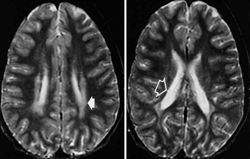
VIsual Difficulties Characteristic of PVL CVI
As indicated above, vision problems can be another result of Periventricular Leukomalacia. This type of visual impairment is known as Cerebral Visual Impairment as its origin is in damage to the cerebral region of the brain during development or birth, in this case, to the inner sub-cortical white matter. Advances in the medical field are enabling doctors to save the lives of more and more premature infants, the frequency of CVI due to PVL is increasing in the developed world. Visual impairment due to PVL CVI ranges from total blindness to near normal vision. It has been found from MRI scans and Teller Acuity Card assessments during studies of these children that the amount of reduction to the white matter tissue near the ventricles corresponds to the degree of impairment. Loss of acuity, limited eye movement ability or motility, reduction to the lower field, and difficulty perceiving motion due to dorsal stream dysfunction are all characteristics frequently present in children with CVI caused by PVL. (Dutton) When comparing children whose CVI results from striate cortex damage versus those whose CVI results from PVL, the greatest difference is in the amount of improvement in visual function seen throughout the child’s development. Those with Non-PVL CVI tend to show significantly greater improvement than those whose visual impairment is rooted in deep white matter damage. Fewer PVL patients show improvement and those who do show it at lower levels.
Why Does It Matter?
As teachers, when considering the causes of visual impairment in children, it is paramount to keep in mind the goal of doing so is to provide interventions and educational supports that are as accessible and uplifting to the children and families we serve. Designing effective instruction requires us to value, respect, and seek to thoroughly understand our students’ challenges as well as hold high expectations for them. This means getting an understanding of how they see and why this is the case from a medical perspective. Only once we understand where our students are coming from can we partner with them to move forward.
References
Cedars Sinai. (n.d.). Periventricular Leukomalacia (PVL) in Children. Retrieved February 5, 2022, from https://www.cedars-sinai.org/health-library/diseases-and-conditions—pediatrics/p/periventricular-leukomalacia-pvl-in-children.html#:~:text=Periventricular%20leukomalacia%20(PVL)%20is%20a,inner%20part%20of%20the%20brain.
The Children’s Hospital of Philadelphia. (2014, August 24). Periventricular leukomalacia (PVL). Children’s Hospital of Philadelphia. Retrieved February 5, 2022, from https://www.chop.edu/conditions-diseases/periventricular-leukomalacia-pvl
Lanzi G;Fazzi E, Uggetti C, Cavallini A, Danova S, Egitto MG, Ginevra OF, Salati R, Bianchi PE, (1998, June 29). Cerebral visual impairment in Periventricular Leukomalacia. Neuropediatrics. Retrieved February 5, 2022, from https://pubmed.ncbi.nlm.nih.gov/9706625/
Lueck, A. H., & Dutton, G. (2015, Ch. 3). Vision and the brain: Understanding cerebral visual impairment in children. AFB Press.
Martín, M. B. C., Santos-Lozano, A., Martín-Hernández, J., López-Miguel, A., Maldonado, M., Baladrón, C., Bauer, C. M., & Merabet, L. B. (2016). Cerebral versus Ocular Visual Impairment: The impact on developmental neuroplasticity. Frontiers. Retrieved January 29, 2022, from https://www.frontiersin.org/articles/10.3389/fpsyg.2016.01958/full
Periventricular leukomalacia (PVL) brain injuries. Periventricular Leukomalacia | PVL Brain Injury – Symptoms, Treatment & Effects. (n.d.). Retrieved February 5, 2022, from https://www.birthinjuryhelpcenter.org/periventricular-leukomalacia-pvl.html
Poinsett, P. M. (Ed.). (2020, June 19). Periventricular leukomalacia (PVL) and Cerebral Palsy. Cerebral Palsy Guidance. Retrieved February 5, 2022, from https://www.cerebralpalsyguidance.com/cerebral-palsy/causes/periventricular-leukomalacia/
U.S. Department of Health and Human Services. (2019, March 27). Periventricular Leukomalacia Information Page. National Institute of Neurological Disorders and Stroke. Retrieved February 5, 2022, from https://www.ninds.nih.gov/Disorders/All-Disorders/Periventricular-Leukomalacia-Information-Page
U.S. National Library of Medicine. (2022, January 12). Periventricular Leukomalacia: Medlineplus medical encyclopedia. MedlinePlus. Retrieved February 5, 2022, from https://medlineplus.gov/ency/article/007232.htm
Warf, B. C. (2012). Periventricular leukomalacia: Boston Children’s Hospital. Boston Childrens Hospital. Retrieved February 5, 2022, from https://www.childrenshospital.org/conditions-and-treatments/conditions/p/periventricular-leukomalacia