
Sclera
Description
The formal name for the white of the eye is the “sclera”. A millimeter thick, disorderly forest of crossing collagen fibers, it encircles the majority of the globe or eyeball like a suit of armor, with only the cornea and optic nerve remaining uncovered. The irregularity of the fibers gives it both its opaque coloring and its strength. Of the entire globe, 5 out of 6 parts are protected by the sclera, with the cornea taking up only 1/6 of the surface area. The sclera is located in the outer layer of the globe and connects to the limbus or border zone of the cornea in the anterior, and to the myelin sheath covering of the optic nerve in the posterior. It is also covered by the conjunctiva when eyes are closed. The sclera is composed of four layers. Going from anterior to posterior, these layers are: the episclera, the stroma, the limina fusca, and the endothelium.
Function
The sclera serves three major functions: protection, structure, and attachment surface. Firstly, its rugged connective tissue shields the internal parts of the eye vital to producing a clear image from external injury. Structurally, the sclera is like a surrounding wall that holds up the shape of the globe. It also supports the maintenance of intraocular pressure by providing resistance for the vitreous and aqueous humors. Thirdly, the sclera is the hard surface that ocular muscles connect to. Think of a surface board in which the muscles that control eye motility are plugged into.
The dynamic nature of the sclera is particularly remarkable. Whether or not it could be considered a primary function of the sclera, this tissue actively adapts to help the rest of the components to form as clear an image as possible. When the retina receives a blurry image, a signal is sent that tells the scleral tissue to expand or contract in a way that nudges the retina into a better position for receiving a clear picture.
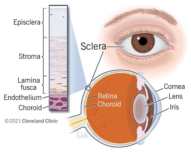
Caption: Diagram shows the sclera wrapping around the vitreous chamber of the eyeball. A close-up illustration shows the four layers of the sclera: episcleral, stroma, lamina fusca, and endothelium. The choroid is right underneath these layers. The opposite end of the diagram identifies the cornea, iris and lens positions.
Conjunctiva
Description
Another protective mechanism of our eyes is the conjunctiva. Essentially, it is a wrapper that fits neatly across and between the sclera and inner eyelid to keep particles from reaching your eye and making their way around the globe to the back as well. This thin but effective membrane is made of three parts: the palpebra is the part lining the inside of the eyelids, the bulbar is the piece that covers the sclera or white of the eye, and then of course there is a piece that connects the two regions known as the fornix. The conjunctiva cannot be easily seen in the mirror as it is clear.
Function
In the same way that an umbrella prevents the majority of rain drops from reaching your head, the three sections of the conjunctiva as a whole are situated in such a way as to block external particles from getting through to the globe itself. In addition to keeping irritants like allergies out and guarding the tissue of the sclera from damage, it also plays a key role in the lubrication process. Our tears have three layers to them and each is vital to their efficacy. The first layer is made by meibomian glands is oily to keep them from evaporating too fast. The watery layer in the middle is made by the lacrimal glands to keep things moist. The conjunctiva is responsible for the third layer which contains mucus to spread the tears around to fully cover the eye. The shape and position of the conjunctiva also helps to hold this moisture in longer. A problem with the conjunctiva can result in an eye infection due to particles getting through to the sclera or to dry eyes due to poor lubrication. For example, conjunctivitis, commonly called Pink Eye, occurs when a bacteria, virus, or allergens infect this part of the optical system. This condition is minor and can be treated with antibiotic eye drops or often goes away on its own.
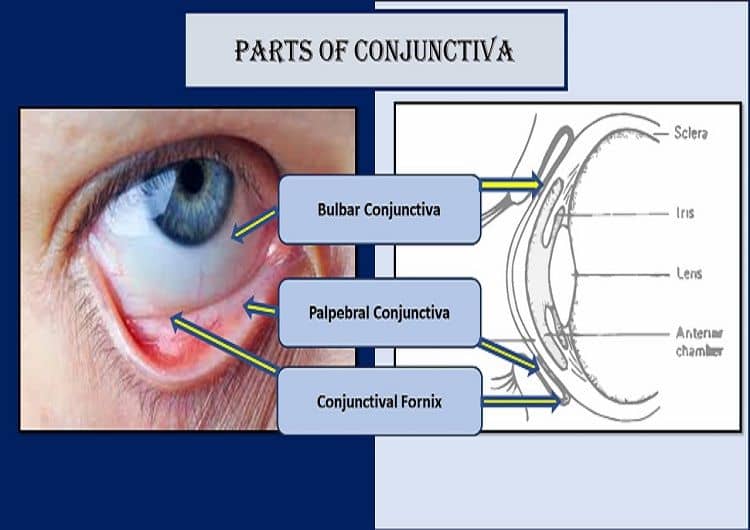
Caption: Figure shows the eye with arrows indicating the areas covered by the conjunctiva. Then on the right side, a magnified view of the conjunctiva is shown with the three sections highlighted: bulbar conjunctiva , palpebral conjunctiva, and conjunctival fornix.
Cornea
Description
Made of the same kind of tissue that makes up our skin, the cornea can be thought of as a transparent dome capping off the visible part of the eye’s camera equipment (iris, pupil, and limbus or the circle of colorful stuff in the center that we see in the mirror). It takes up 1/6 of the eye’s surface area with the sclera taking the other 5/6. As it is clear, this powerful outer structure, enables light to flow into the eye for processing. Because it is transparent, it is only noticeable when light reflects off of it and is much wider than it is thick with approximately 12 mm of width to less than 1 mm of depth. The cornea is comprised of five layers. From anterior to posterior, these are: the epithelium, Bowman’s membrane, stroma, Descemet membrane, and endothelium. The stroma is the thickest and the endothelium the thinnest. The surface of the cornea is covered in a tear film containing lipid, aqueous, and mucin layers that are necessary for maintaining its health.
Function
The choroid serves as a lifeline that shuttles oxygen and nutrients to the retina. When this vital function is not working effectively, deterioration to the macula and/or retina can be the result. Secondarily, this structure helps the retina maintain the correct temperature by a cooling or warming blood flow as needed. It also aids in drainage to maintain intraocular pressure.
Another amazing thing about the choroid is that the melanin pigment in it helps to prolong our vision by absorbing harmful light rays, thus limiting how many make it through to the retina. This pigment also serves to protect the blood vessels from contamination. However, the melanin does cause the red eye effect that is so unpleasant to see in flash photography.

Caption: A diagram depicts a view of the corneal layers as seen from a microscope. The layers are: epithelium or surface layer, stroma or structural layer, and endothelium or pump layer.
Choroid
Description
The choroid is both the largest and most posterior component of the Uveal tract or middle vascular layer of the eye that includes the structures underneath the cornea but above the lens (Iris, ciliary body, and choroid). It is primarily composed of blood vessels. Think of the choroid as a thick tube with many smaller tubes running through it to the outer retina. The choroid is located between the sclera and retina and in length, runs from the ora serrata to the optic nerve. It is called the “vascular layer” because of its many blood vessels and capillaries that convey nutrients. In addition, it also contains pigment and epithelial cells. The choroid itself contains four layers, from anterior to posterior, these are the suprachoroid lamina, choroidal stroma, choriocapillaris, and Bruch membrane.
Function
The choroid serves as a lifeline that shuttles oxygen and nutrients to the retina. When this vital function is not working effectively, deterioration to the macula and/or retina can be the result. Secondarily, this structure helps the retina maintain the correct temperature by a cooling or warming blood flow as needed. It also aids in drainage to maintain intraocular pressure.
Another amazing thing about the choroid is that the melanin pigment in it helps to prolong our vision by absorbing harmful light rays, thus limiting how many make it through to the retina. This pigment also serves to protect the blood vessels from contamination. However, the melanin does cause the red eye effect that is so unpleasant to see in flash photography.

Caption: The vascular choroid layers flowing near the retina
Ciliary Body
Description
The ciliary body is a ring-shaped tissue starting at the base of the iris at the back of the middle layer that separates the anterior and posterior chambers. It has two main parts: the ciliary muscle and the ciliary process. Think of the ciliary muscle as two arms that are attached to either side of the lens via zonules that extend like fingers, holding them in position on the lens capsule. The ciliary muscles exert force to push and pull the lens into the right shape for accommodation to take place. The ciliary process manufactures the aqueous humor that flows between the anterior and posterior chambers by way of the pupil. The ciliary body is located between the iris and the lens up against the sclera. It is made of smooth muscle, connective, and vascular tissues and comprises the following layers: supraciliaris, ciliary muscle, ciliary stroma, and two layers of ciliary epithelium.
Function
The ciliary body has two main functions: 1) to change the chape of the lens allowing accommodation to occur, and 2) to produce the aqueous fluid that nourishes the structures of the anterior chamber such as the cornea. The ciliary muscle is necessary for accommodation, which happens when the lens contracts to bring objects into focus on the retina when the target is suddenly brought into closer range from a distance. The ciliary process contained in the vascular, stromal, and epithelial layers, has its own job of making aqueous fluid, then secreting it into the posterior chamber. The three stages of this process are called diffusion, ultrafiltration, and active secretion.
In short, the ciliary body is vital for clear perception of near objects. For example, enabling us to read. And just as importantly, the ciliary body keeps the eye healthy, helps it retain its full shape, and maintains pressure.

Caption: Diagram of the vascular and neural layers of the eye shows the Uveal tract with labels indicating the ciliary region, iris, sclera, nerves, and choroidal veins.,
Iris
Description
The Iris is the colored ring in your eye. The amount of pigment in it melanin determines your eye color. A lot of pigment means brown eyes, a little means blue, and somewhere in the middle results in the many varieties and shades that make all our eyes unique. The iris sits underneath the cornea, perched above the lens and surrounding the black hole (pupil) in its center. It is part of the Uveal tract in the middle vascular layer of the eye. The iris is composed of nerves, muscle, and aqueous humor.
Function
The primary function of the iris is to regulate the amount of light that can enter the eye. The nerves in the iris sense how much light there is and signal the muscles to respond accordingly. The muscles are used for changing the shape of the pupil in response to the amount of light present. They contract to make the pupil smaller in bright places. This prevents too much light from reaching the retina at the back, which would lead to damage and blurring. These muscles dilate to enlarge the pupil in dim places to allow you to see when there is little light present. The ability to adjust and see when going from light to dark places and vice versa is called adaptation. The iris’s response to changes in light levels, called pupillary light reflex, is called sympathetic because it is automatic, not requiring effort on our part to occur.
The ability to alter the size of the pupil is very important for maintaining clear vision and limiting sun damage. Aniridia is a condition where a person is born without an iris. Without it, too much light pours into the back of the eye through the pupil causing very blurred vision and exposing the delicate inner structures to more harmful light rays.


Caption: Close up view of an iris with the pupil shown in the center. Labeled regions include the folds of the iris, pupillary margin, anterior surface, inner border of iris, ciliary margin, outer border of the iris.
Aqueous Humor
Description
Both the anterior and posterior chambers contain a clear, watery fluid called aqueous humor. The anterior chamber is the region from the cornea to the iris, while the posterior is from the iris to the lens. Aqueous is secreted from the ciliary body into the posterior chamber. Then it flows between the iris and lens and through the pupil into the anterior chamber. After circling the anterior chamber, it exits via the sponge-like trabecular meshwork and then the tube-like canal of schlemm, eventually filtering into the circulatory system. Aqueous is 99.9% water, with only a tiny fraction containing important nutrients.
Function
The continuous cycle of production, circulation, and drainage of aqueous throughout the posterior and anterior chambers of the eye contributes to its health in three main ways. Firstly, this process delivers needed nutrients that keep the eye functioning. For example, it brings oxygen and glucose to the cornea and lens. Secondly, it washes waste particles out of the eye. Thirdly, a consistent volume of it maintains the interocular pressure in the front areas of the eye. Balancing the rate of secretion with the rate of drainage is crucial for a stable, healthy pressure.
When the flow in and flow out amounts are not equal, the pressure grows too high, resulting in ocular tissue deterioration. This is known as glaucoma. Rising of interocular pressure is caused by either the over production of aqueous humor or a blockage that prevents it from draining out effectively. This is also called ocular hypertension and, if unchecked, leads to glaucoma which can result in blindness.

Caption: Figure shows the areas in the eye where aqueous humor is produced, using labeled arrows to indicate where parts of the process occur, including where it forms, flows through, and exits out of the eye.
Canal of Schlemm
Description
The canal of Schlemm is a circular pipe running through the perimeter region between the cornea and sclera called the limbus. It is a vein that flows with aqueous rather than blood. Aqueous humor filters into it from the trabecular meshwork and is then conducted out of the eye’s anterior chamber.
Function
The canal of Schlemm is the means by which aqueous fluid exits the eye, allowing interocular pressure to be maintained as long as the rate of aqueous going out stays consistent with the amount being produced in the ciliary body process.

Caption: Diagram shows the movement of Aqueous Humor out of the eye via the canal of Schlemm.
Lens
Description
The lens we are born with is called the crystalline lens, as opposed to a surgically implanted IOL (interocular lens). This structure sits behind the iris in a capsule that is like a bag in between the back of the posterior chamber and the front of the vitreous chamber. The lens is the size of a skittle or about 10mm long. It is held in place by suspensory ligaments called zonulas that are pushed and pulled on by the ciliary muscle to shape the lens into that needed for refraction of light to be focused through to the retina.
The natural lens is made of translucent fibers that enable it to be flexible and clear so it can change it shape while light passes through it. The crystalline lens contains four layers: capsule, epithelium, cortex, and nucleus.
Function
The essential function of the crystalline lens is to refract and then transmit light rays from the front chambers of the eye (anterior and posterior) through to the retina at the back. While the cornea does do the bulk of refraction, the lens still handles a third of this job, ensuring a clear picture can be sent to the retina. This focusing ability called refraction happens as the lens thickens and thins depending on the distance of the target object. This adjustment to distances is called the process of accommodation. A thinner, relaxed lens is focusing on a more distant target, while a condensed, thicker lens is straining to focus on a nearby target at close range. Without the lens’ ability to accommodate, our eyes would not be able to pick up details at varying distances and focusing on a near target would be much harder.

Caption: Figure illustrates the effect of the crystalline lens on image quality focused on the retina. The lens is shown between the ciliary body and zonulas. Three areas of the lens are labeled from top to bottom: lens epithelium, lens capsule, and lens fibers.
Suspensory Ligaments (Zonules)
Description
Also called zonules, the suspensory ligaments are fastened to the lens capsule on one end and the ciliary body muscle on the other. The ciliary muscle expands and contracts to push and pull on these ligaments, which in turn, cause the lens to change shape. These miniscule fibers are needed to fix the lens in place.
Function
The suspensory ligaments or zonules are a key feature in the accommodation process. They are the means by which the ciliary muscle controls the shape of the lens so that near and distant objects can be focused properly and refracted to the retina.

Caption: A model of the posterior chamber indicates the lens, ciliary muscles, and zonules.
Pupil
Description
The pupil is the black hole of emptiness at the center of the eye in the anterior chamber that allow slight to pass through into the back of the eye where images are processed in the retina. While we think of the pupil as the black round structure at the very center of the eye, it is actually open space in the middle of the iris. The pupil looks black because light passes through it and is absorbed by the retina, rather than reflecting off of anything. However, in flash photography, pupils can appear red. This is because light is reflecting off the red retina. The pupil in the human eye is approximately 2 to 8 millimeters but can vary. It grows larger in dim lighting and shrinks in bright settings. The pupil tends to get smaller with age.
Function
The iris and pupil work together to regulate how much light enters the eye. The pupil is surrounded by the iris which has muscles to either constrict or dilate, causing the pupil opening to grow or shrink in size. The pupil is dilated when there is not a lot of light available to make best use of what there is, allowing night vision to occur. In bright light, the pupil constricts to protect the back of the eye from being exposed to too much light. If the amount of light flooding into the eye was not regulated, the result would be glare, pain, and damage to the retinal tissue. Because of the accommodative pupillary response, pupils also constrict somewhat when the eye focuses on a near object.

Caption: Diagram shows the working of the iris to control the size of the pupil. When iris muscles are constricted, the pupil is small. When the iris muscles are dilated, the pupil is large. The iris has a dilator radial muscle and a sphincter circular muscle.
Retina
Description
The retina is the light processing center where light rays are received and translated into electrical impulses, then sent on to the brain. The retina lines the whole back wall of the eye and is attached to the optic disk to which the optic nerve is connected. It consists of layers of neural tissue and light sensitive cells called photoreceptors that come in rod and cone varieties. Rods are for night vision and cones are for daylight vision. They are what creates the electrical signals sent to the brain. In addition to the photoreceptors, the retina contains the macula, fovea, and peripheral retina. Many rod cells are in the peripheral retina whereas cone cells are in the central macula area.
Function
The retina converts the light that enters the eye into visual images that can be sent to the brain for further processing. It is where both peripheral and central vision are made possible. The peripheral retina contains nerves and rods that allow for peripheral and night vision. The central retina contains cones that allow for detailed central vision in daylight. The retina is like a bridge between the eyes and the brain. The retina creates the electrical signals that the brain can then interpret as images.

Caption: Diagram of the retina with important features labeled including: optic disk, central retinal artery, retinal venules, retinal arterioles, macula, and fovea.
Fovea Centralis
Description
The fovea is a tiny divot at the very center of the macula which in turn is at the center of the retina. It provides the sharp, detailed vision in the central two degrees of our field of view. This one little spot where we see the clearest and most detailed is also called foveal vision. The fovea is about 1.5 mm in diameter and packed with 199,000 cone photoreceptors every square mm. There is a pit in the middle of the fovea called the foveola. There are no blood vessels in the fovea because it receives it blood supply from the choroid.
Function
The function of the fovea is to provide high acuity central vision. The fovea gives us the small area of maximum acuity vision with the sharpest clarity of detail. Both the high density of cones and the lack of blood vessels contribute to seeing clear perception of colors and fine details.

Caption: Fovea centralis indicated as a circle in the center of the fovea in the middle of the macula.
Macula
Description
The macula is the central region of the retina that contains many photoreceptors to provide detailed central vision. It is oval shaped, yellow in color, and about 5 mm in diameter. The macula has six layers: the umbo, foveola, foveal avascular zone, fovea, parafovea, and perifovea.
Function
The macula’s primary function is central vision, as well as color vision and seeing fine details. Secondarily, the yellow pigment also works to absorb unhealthy blue and ultraviolet light to protect the surrounding retina.

Caption: Close up of the macula at the center of the retina.
Vitreous
Description
The vitreous humor is the gel-like substance that fills the vitreous chamber located between the lens at the back of the posterior chamber and the retina at the back of the eye which it attaches to. The vitreous chamber is the largest structure in the globe, taking up 80% of the space. Vitreous humor is thicker and less watery than aqueous humor. It is transparent so that light can pass through it. Although vitreous is comprised mainly of water, it also has other particles including collagen, proteins, electrolytes, and glycosaminoglycan sugars. It contains antioxidants that support the health of the lens. The vitreous gel enables oxygen and other nutrients to be transported to the back of the eye from the front. In order to stay transparent, it does not contain any blood vessels.
Function
The central function of the vitreous chamber and humor is to maintain the shape of the eye and the pressure that keeps it sturdy. The clear, watery nature of the humor is necessary for allowing light to reach the retina for processing. Clear central vision is possible because of light going through a clear vitreous and being focused on the central retina called the macula. A third function of the vitreous is shock absorption such as those caused from head movements, exercise, sneezing, or even a head injury. The vitreous helps to minimize any damage done by these movements to the internal structures at the back of the eye.

Caption: Diagram of the eye indicates the vitreous chamber as the largest central area of the globe. The anterior and posterior chambers are also labeled and shown as much smaller areas at the front.
Ora Serrata
Description
The ora serrata is the name given to the junction or border region between the choroid and the ciliary body. It surrounds the retina at the back of the eye. It is serrated and jagged in shape. It is where the ciliary body ends and transitions to the photoreceptor rich retinal layers. It is about 2 mm in width and contains projections of retinal tissue.
Function
Although information on the function of the ora serrata is quite scarce, it helps to hold the retina in place and protects it from detachment. It also supports peripheral vision by protecting the inner retinal tissue.

Caption: Magnified view sketch of the ora serrata retinal border region.
Optic Nerve
Description
The optic nerve can be thought of as the cable that connects the eyes to the brain. It starts out as small fibers in the ganglion cells in the retinal interior (called the nerve fiber layer). These strands grow to millions and bunch up into a bubble shape at the back of the eye which is referred to as the optic nerve. This nerve reaches all the way to the visual system in the brain and connects to the optic chiasm. The optic nerve is 5 or more cm long and is comprised of four segments lengthwise: intraocular, intraorbital, intracanalicular, and intracranial. Another name for the optic nerve is the second cranial nerve as it is an extension of the brain. The many nerve cells bundled together transmits electrical signals from the eyes to the brain so that vision can form. The optic nerve’s blood supply is provided by a branch of the internal carotid artery.
Function
The optic nerve’s primary duty is plugging the eye into the brain. The eye might collect the sensory information for vision, but it is the brain that does the heavy lifting of processing the input into a meaningful and understandable image. This requires that all information collected by the eyes be translated into electrical impulses the brain can interpret. The translation from light to electrical impulses takes place in the retina, then those signals are sent to the optic nerve connected to the back of the retina where they travel along the optic nerve, meeting up with the opt8ic chiasm along the way, and continuing up the optic nerve all the way to the visual system in the brain along. Secondarily, the optic nerve also enables color, brightness, and contrast perception. What is more, the optic nerve is the reason for the neurological reflexes of light and accommodation. The light reflex is why the pupils constrict when bright light shines on one or both eyes. The accommodation reflex lets the eyes adjust for near vision by helping the lens thicken, although accommodation is primarily a function of the ciliary body, suspensory ligaments, and lens.

Caption: Photograph of the optic nerve shown at the back of the retina.
Optic Disk (Blind Spot)
Description
The optic disk, also called the optic nerve head, is a circular, raised structure at the back of the eye behind the retina, specifically between the macula and the peripheral region. It is typically pink, orange, or yellowish but can appear pale or white when less healthy. Because there are no photoreceptor cells (rods or cones) on the optic disk, it is referred to as the physiologic blind spot. This disk is the point where the retina and optic nerve are linked. It is also the place where the main artery that supports the retina comes into the eye. At the center of the optic disk there is an indent known as the physiologic cup where the optic nerve joins to the disk and subsequently the retina.
Function
The function of the optic disk is to indicate the beginning of the optic nerve region as it connects the retina to the optic nerve so that sensory signals can be sent to the brain. It also contains the vein that supplies blood to the retina. This nerve head is where the optic nerve enters the eye. Ganglion cell axons exiting the retina join to it so electrical signals run smoothly from the retina to the optic nerve and up to the brain.

Caption: The optic disk is a round section at the back of the eye with no photoreceptors where the retina and optic nerve are connected.

Caption: Diagram shows the location of the optic disk as a circled area at the back of the eye.
References
Ayaga, V. (2023, August 29). Retina: Anatomy, functions, and Conditions. Vision Center. https://www.visioncenter.org/eye-anatomy/retina/
Bedinghaus, T. (2022, August 11). What is the choroid?. Verywell Health. https://www.verywellhealth.com/choroid-eye-anatomy-choroid-definition-3421675
Belden, S. (2023, January 17). Optic disc. All About Vision. https://www.allaboutvision.com/eye-care/eye-anatomy/optic-disc/
Blackwell, C. (2014, March 10). Cornea 1: A remarkable structure. YouTube. https://youtu.be/Xf21nk2w8g0?si=0PmTzOiU-SxZi7i8
Cassel, G. H. (2021). The eye book: A complete guide to eye disorders and health. Johns Hopkins University Press.
Cassin, B., & Rubin, M. L. (2012). Dictionary of eye terminology. Triad Pub.
Porter, D. (2021, May 28). Zonules. American Academy of Ophthalmology. https://www.aao.org/eye-health/anatomy/zonules
professional, C. C. medical. (n.d.). Conjunctiva: Anatomy, function & common conditions. Cleveland Clinic. https://my.clevelandclinic.org/health/body/24329-conjunctiva
Remington, L. A., & Goodwin, D. (2022). Clinical anatomy and physiology of the visual system. Elsevier.
Shinbashi, M., Randolph, J., & Bhagat, N. (2023, April 11). Ora Serrata. EyeWiki. https://eyewiki.aao.org/Ora_Serrata
Sprabary, A. (2021a, March 2). Aqueous humor. All About Vision. https://www.allaboutvision.com/eye-care/eye-anatomy/aqueous-humor/
Sprabary, A. (2021b, March 10). Vitreous humor: Gel of the eye. All About Vision. https://www.allaboutvision.com/eye-care/eye-anatomy/vitreous-body/
Troy Bedinghaus, O. (2023, July 3). Learn about the anatomy of the macula of the eye. Verywell Health. https://www.verywellhealth.com/macula-anatomy-function-and-significance-4771995